|
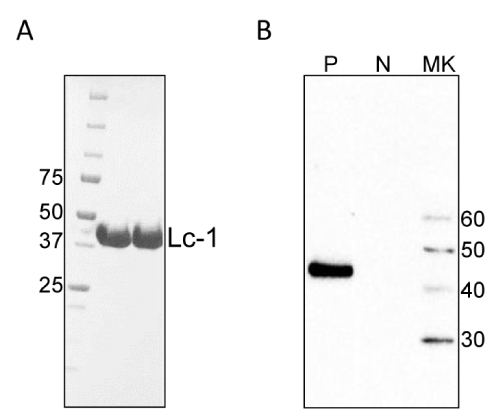
| Figure 2: The r56Lc-1 protein after purification. (A) Two different pools of purified Lc-1 at 10 μg per lane were loaded on to 4-20% gradient SDS-PAGE (Bio-Rad). After gel electrophoresis, the gel was stained with Gelcode Blue (Pierce) per manufacturer's instruction. The location of Lc-1 is shown as indicated between 37 kDa and 50 kDa MW markers (B) Purified r56Lc-1 was evaluated to determine reactivity with serum samples collected from mice infected by Lc-1 chigger (P) or naïve chigger (N) as shown on the western blot. MK: molecular weight marker (kDa). |