|
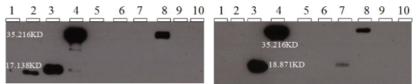
| Figure 2: Western blotting detection of GnRH-I fusion proteinobtained from transfected COS 1 cells and labelled with anti-GnRH-I (left) and anti V5 antibodies (right). The first [10], second[6] and the new vaccines were used to transfect COS 1 cells. Lane 1shows the Kaleidoscope ladder, lanes 2, 3 and 4 containingtransfected COS1 cell lysate of the first, second and new vaccinesrespectively. Lanes 6, 7 and 8 containing transfected COS1 cellculture supernatant of the first, second and new vaccine repectively.Lane 5 and 9 containing non-transfected COS1 cell lysate andculture supernatant respectively (negative control) and lane 10lacking both anti-GnRH-I (left) and anti V5 antibodies (right) inthe Western blotting detection of the fusion protein. The newlyengineered vaccine produce fusion protein (35.216 KD) detectedboth in cell lysate (lane 4) and cell culture supernatant (lane 8).GnRH-I fusion protein (17.138 KD) of the first vaccine (lane 2) wasdetected in the cell lysate (lane 6). GnRH-I fusion protein (18.871KD) of the second vaccine was detected both in cell lysate (lane 3)and cell culture supernatant (lane 7). GnRH-I fusion protein wasnot detected in any of the control lane 5, 9 and 10. |