|
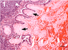
| Figure 1: Mesenteric lymph node, deep cortex. A columnar epithelium with numerous interspersed goblet cells (*) that secret an eosinophilic material (black arrows) is present. At the right side of the image, a strong eosinophilic exudate with numerous neutrophils can be observed. A thin eosinophilic structure similar to a muscularis mucosae layer can be observed under the epithelium (white arrow). Hematoxylin/Eosin staining, Scale bar: 100 μm. |