|
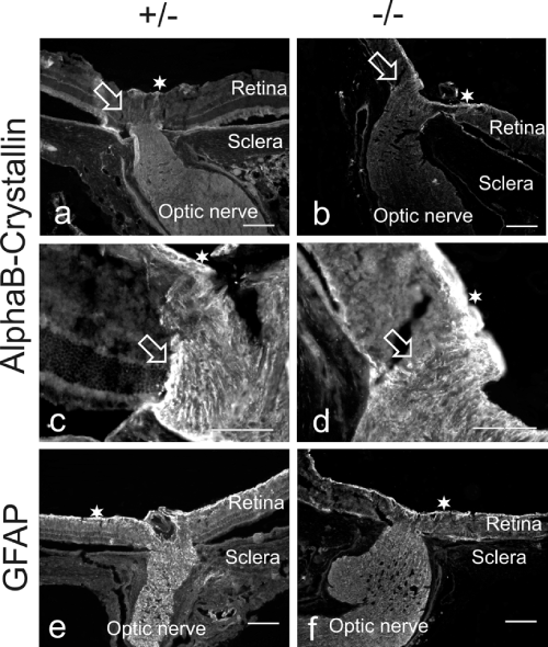
| Figure 1: Micrographs through the optic nerve head of healthy (+/-) and dystrophic (-/-) RCS rat eyes (both 12 months of age), stained for alphaBcrystallin and GFAP. The zone between the retina and the optic nerve head is marked with an open arrow. A star marks the inner retinal layer. A mild increase of alphaB-crystallin is only seen in the innermost layer of the optic nerve head (stars). Scale bar equals 100 μm. |