|
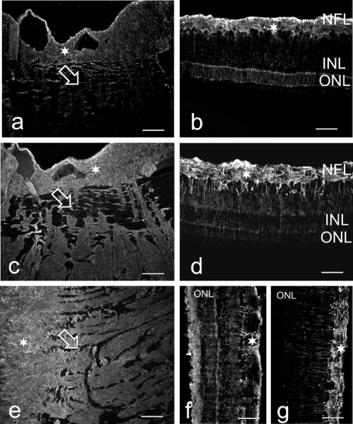
| Figure 3: Micrographs through the optic nerve head (a,c,e; scale bar 200 μm) and central retina (b,d,f,g; scale bar 100 μm) of the porcine (a-d) and bovine (e-g) eye, stained for alphaB-crystallin (a,b,e,f) and GFAP (c,d,g). Astrocytes in the lamina cribrosa (open arrow) and in the prelaminar region (stars in a,c,e) as well as in the nerve fiber layer of the retina (stars in b,d,f,g) staine positive for alphaB-crystallin. Single Muller cells show also alphaB-crystallin immunoreactivity, but less regular than GFAP (parallel stained lines through the retina in d,g). NFL=nerve fiber layer, INL=inner nuclear layer, ONL=outer nuclear layer. |