|
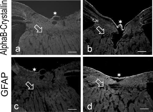
| Figure 4: Micrographs through the optic nerve head of a healthy (a,c) and a glaucomatous (b,d) monkey eye (one animal, 12 years of age), stained for alphaB-crystallin and GFAP. The lamina cribrosa region (open arrow) and prelaminar region showed no staining difference between glaucoma and control for alphaB-crystallin, but a clear increase in GFAP. Only the innermost line of the optic nerve head (star) showed an increase in alphaB-crystallin staining. Scale bar equals 200 μm. |