|
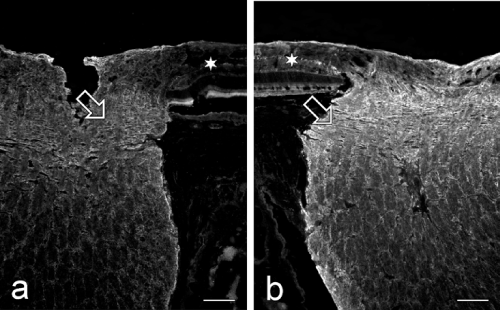
| Figure 5: Micrographs through the optic nerve head of a healthy (a) and a dystrophic (stage 2, b) Abyssinian cat eye (both animals 4 years of age), stained for alphaB-crystallin. The homogenous staining in the normal eye (lamina cribrosa as indicated by the open arrow, prelaminar region, rim of the optic nerve bundles) is somewhat increased in the dystrophic eye. A star marks the retina which shows alphaB-crystallin staining in the nerve fiber layer. Scale bar equals 200 μm. |