|
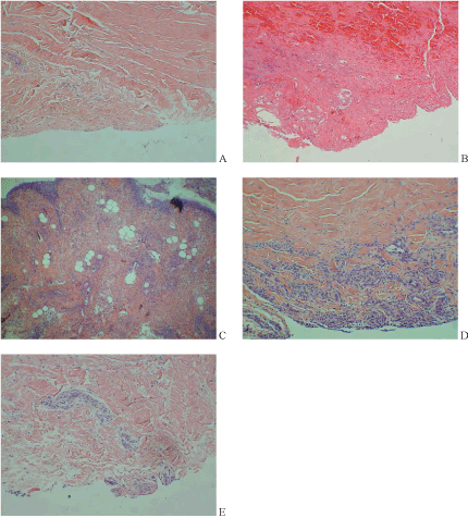
| Figure 1: Histology slides of middle carpal joint synovium taken at post mortem evaluation of each study horse. All images were stained with H&E and taken at 40x magnification. A) Horse 1, control sample (right middle carpal synovium). B) Horse 1, treated sample (left middle carpal synovium). Note the obvious presence of red blood cells, lack of collagen fibre orientation, and small cell infiltrate within the fibrolayer of the synovium. The subsynovial layer appears to be detached or absent, most likely consistent with handling artifact. C) Horse 2, treated sample (left middle carpal synovium). Note increased cellularity in this sample as compared to A, however, in this animal’s self-controlled sample (right middle carpal synovium), mild hypercellularity was also present. D) Horse 3, treated sample (left middle carpal synovium). Note the inflammatory infiltrate seen in the subsynovia. E) Horse 4, treated sample (left middle carpal synovium). Note the small amount of inflammatory cell infiltrate accompanied by mild disorganisation of collagen fibres. |