|
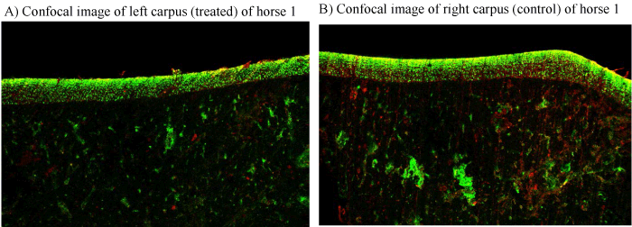
| Figure 2: Confocal microscopy of Horse 1. Figure1A is the treated, left proximal third carpal bone sample including articular cartilage and subchondral bone within the left middle carpal joint. Figure 1B is the control, right proximal third carpal bone sample. No appreciable differences were present between treated and control samples on confocal microscopy. |