|
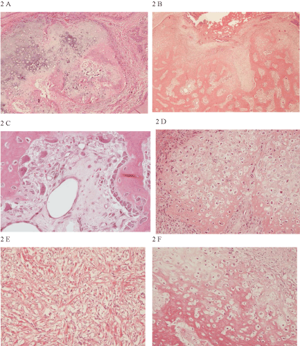
| Figure 2: Primary malignant mixed mammary tumour demonstrating cartilage (A), (objective x 10) and bone tissue (B), (objective x 4). Low-grade bone tissue with osteoblasts lining the matrix to the right (arrow) and multinucleated osteoclasts to the left (*), (C) as well as less differentiated cartilage with single chondrocytes in the lacunas in the matrix (D), (objectives x 40 and x 20). Two years after surgical removal of this primary tumour (in gland R3) the dog was post-mortem examined and a spindle cell tumour was found in gland R4 (E) as well as a combined osteosarcoma in gland R5 (F), (objectives x 20). Light micrographs from dog No. 117 with haematoxylin and eosin (HE) staining. |