|
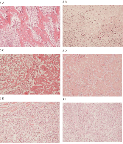
| Figure 5: Primary low-grade combined mammary osteosarcoma with presence of neoplastic osteoblasts and an osteoid matrix (A). Cartilage was found only in a minor part of the primary tumour (B). The lung metastases were low-grade and composed of bone (C and D). Metastases in the pleura, myocardium and kidneys had a similar morphology (not shown) whereas the metastases in the diaphragm were high-grade (E). Probable metastases in the spleen showed spindle shaped cells that formed a whirl-like growth pattern (F). Light micrographs of dog No. 144 using objectives x 20 of haematoxylin and eosin (HE) stained sections. |