 |
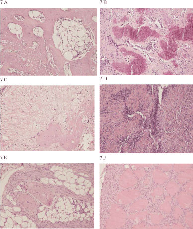
| Figure 7: Light micrographs of tumours formed in nude mice by a cloned cell line established from dog No. 353. A low-grade osteosarcoma with abundant presence of bone matrix and few tumour cells, also an area of suspected bone marrow-like cells (*) were seen (No. 1-3), (A). A high-grade osteosarcoma from the same clone with a large number of tumour cells and less matrix is shown (No. 1-5), (B). Tumours formed by clone No.2 were low-grade to high-grade and as shown in panel C, a calcified (deep purple) bone matrix was seen in the low-grade part of the formed osteosarcoma (No. 2-1), (C). In another tumour formed by the same clone, lowgrade bone tissue was seen in the centre whereas a loose matrix formed the periphery of the tumour (No. 2-2), (D). One of the clones did not form bone tissue in the mice but formed highgrade spindle-cell tumours. These tumours grew very invasive and infiltrated the surrounding tissues such as fat tissue, peripheral nerves (E) and the skeletal muscles (No. 6-1B), (F). Objectives x 20 of haematoxylin and eosin(HE) stained sections. |