|
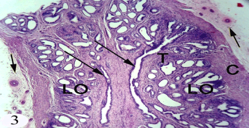
| Figure 3: A photomicrograph of the longitudinal section of the immature seminal gland showing highly vascularized loose connective tissue enveloped the gland (short arrow), capsule (C ), trabeculae (T), small lobules (LO) and central cistern (long arrow). Stain: H& E Obj.x4: Oc.x10 |