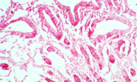
Figure 1:
Small intestine from a calf. Villi and crypts of lieburkuhn are widely separated from each other and mild leucocytic infilteration and edema are seen in the lamina properia (X-100).