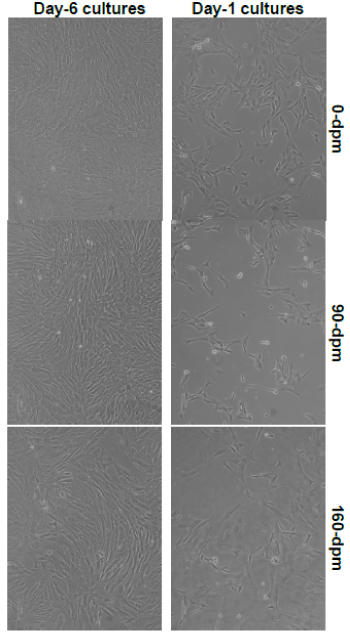
Figure 2:
Comparative fibroblast cell morphology of 3 cell lines (passage 4): Light microscopy, x100 magnification. TS100 inverted microscope and DSL2 camera (Nikon) were used to capture images