|
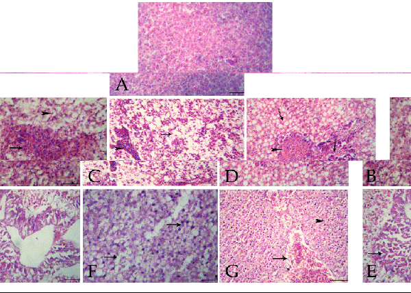
| Figure 1: A: Section of O.niloticus liver showing normal hepatocyte and sinusoidal architectures, H&E (Bar=100 μm). B: Section of O.niloticus liver of second group showing focal area of necrosis infiltrated with numerous lymphocytes and few erythrocytes (arrow) and severe hydropic degeneration (arrowhead), H&E (Bar=100 μm). C: Section of O.niloticus liver of second group showing diffuse hydropic degenerations and vacuolations in the hepatocytes (arrow), and lymphocytes infiltrations in the portal area (arrowhead), H&E (Bar=100 μm). D: Section of O.niloticus liver of third group showing severe congestion of the hepatic blood vessels (arrowhead), hemorrhages (arrow) and diffuse fatty change (irregular arrow), H&E (Bar=100 μm). E: Section of O.niloticus liver of third group showing coagulative necrosis in the pancreatic acini (arrow), H&E (Bar=100 μm). F: Section of O.niloticus liver of fourth group showing moderate fatty change and hydropic degeneration (arrows), H&E (Bar=100 μm). G: Section of O.niloticus liver of fourth group showing severe congestion and hemorrhages (arrow) besides hydropic degeneration (arrowhead), H&E (Bar=100 μm). |