|
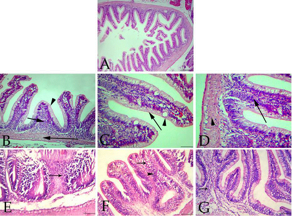
| Figure 3: A: Section of O.niloticus intestine of control group showing normal mucosa, submucosa and muscularis, H&E (Bar=100 μm). B: Section of O.niloticus intestine of control group showing normal mucosa (arrow head), normal submucosa (short arrow) and normal muscularis (long arrow), H&E (Bar=100 μm). C: Section of O.niloticus intestine of control group showing normal lining epithelium of simple columnar cells (arrow) with scattered goblet cells (arrow head), H&E (Bar=100 μm). D: Section of O.niloticus intestine of control group showing normal submucosa (arrow) and normal muscularis (arrow head), HE (Bar=100 μm). E: Section of O.niloticus intestine of second group showing severe necrosis in the intestinal villi and lymphocytes and macrophages infiltrations (arrow), H&E (Ba =100 μm). F: Section of O.niloticus intestine of third group showing intact mucosa with moderate mucinous degeneration (arrow) and few lymphocytes in the submucosa (arrowhead), H&E (Bar=100 μm). G: Section of O.niloticus intestine of fourth group showing mild edema and few lymphocytes infiltrations in the submucosa (arrow), H&E (Bar=100 μm). |