|
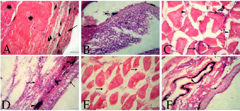
| Figure 4: A: Section of O.niloticus skin of control group showing normal epidermis (arrow), dermis (arrow head) and dermal skeletal muscles (star), H&E (Bar=100 μm). B: Section of O.niloticus skin of second group showing intact epidermis with severe proliferation of epidermal cells, spongiosis and hydropic degeneration (arrows) besides few melanin carrying cells (arrowhead),H&E (Bar=100 μm). C: Section of O.niloticus skin of second group showing Zenker’s necrosis infiltrated with numerous round cells (arrows) and edema (arrowheads), H&E (Bar=100 μm). D: Section of O.niloticus skin of third group showing increased of mucous cells (arrow) and slight vacuolations of the epidermal cells. Edema and aggregations of the leukocytes (irregular arrow) and melanin-carrying cells (arrowhead) were seen in the dermis, H&E (Bar=100 μm). E: Section of O.niloticus skin of third group showing edema and focal hyaline degeneration in the skeletal muscles (arrows), H&E (Bar=100 μm). F: Section of O.niloticus skin of fourth group showing intact epidermis (arrow) with activation of melanomacrophages in the dermis (arrowhead), H&E (Bar=100 μm). |