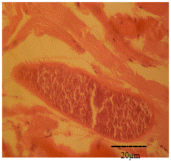
Figure 7:
Photomicrograph showing spiny cyst wall of Sarcosystis in skeletal muscles of Camelus dromedarius stained with H & E. x bar=20 μm.