|
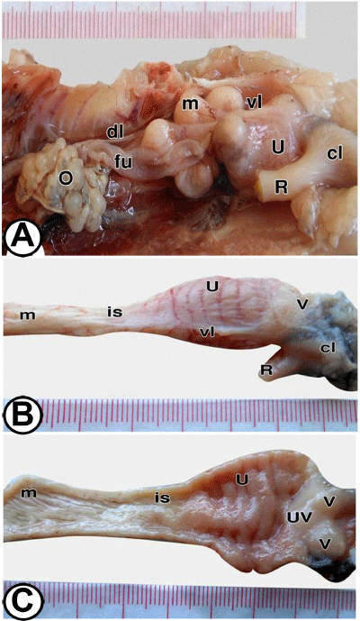
| Figure 2: Gross morphology of the caudal part of the oviduct at the differentiated stage: Figure 2A: at 30-days old, the ovary (O) showing moderately developed follicles, and the oviduct differentiated into infundibulum with its distinct funnel (fu), magnum (m) with many coils that affected by dorsal and ventral ligaments (dl, vl) and uterus (U) that showing oblique ridges externally. cl: Cloaca; R: Rectum. Figure 2B: Higher magnification of the caudal part of the oviduct at 35 days, showing the transverse ridges on the wall of the uterus (U), and the termination of the ventral ligament of the oviduct (vl). Note the junction of the uterus with vagina (v) and isthmus (is) that connected with magnum (m). cl: Cloaca; R: Rectum. Figure 2C: Opened caudal part of the oviduct at 40- days showing spirally twisted folds of the magnum (m), short folds of the isthmus (is), transverse oriented ridges of opened uterus (U), utrovaginal junction (UV) and vagina (V) that showing vaginal sphincter. |