 |
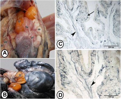
| Figure 3: Gross morphology of the uterus at adult stage and quail samples injected by serum Indian ink. Figure 3A: adult stage showing large mature ovum (O), which is engulfed by funnel part of infundibulum (fu), coils of magnum (m), large darkly pigmented uterus (U) and ventral ligament of the oviduct (vl) attached to the infundibulum, magnum and uterus. Pe: peritoneum; R: Rectum. Figure 3B: Adult quail injected with serum Indian ink showing ovary (O) with blood vessels filled with stain, magnum (m) with stained blood vessels in its wall. Uterus (U) contains an egg and its wall is stained black by serum Indian ink. R: Rectum; vl: Ventral ligament of the oviduct. Figures 3C and 3D: histological specimens injected by serum Indian ink showing peripheral arterial branch (arrow), which supplied clusters of uterine glands. Central arteriole (arrowheads) was found in the bulbs of the primary and secondary folds. |