 |
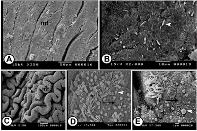
| Figure 7: SEM of the caudal part of the oviduct at undifferentiated and differentiated stage. Figure 7A: The mucosa of the caudal part at 10 days showing longitudinal mucosal folds (mf) separated by deep furrows. Figure 7B: High magnification of the mucosa of the caudal part at 10-days, showing scattered single cilium (arrowhead) among the dome-shaped surface epithelial cells (arrow). Figure 7C: The uterus at 30-days showing wavy parallel longitudinal mucosal folds (mf) separated by deep furrows. Figures 7D and 7E: Higher magnifications of the luminal surface of the uterus at 30-days, showing patches of long cilia (asterisks) and the dome-shaped non ciliated cells with microvilli (arrows). Notice numerous blebs like secretions accumulate on the luminal surface (arrowheads). |