|
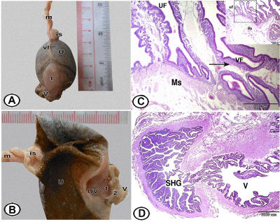
| Figure 9: Gross morphology and histology of sperm host gland in adult quail. Figure 9A: Ventral view of the caudal part of the left oviduct showing the sperm host gland (1) located between vagina (V) and uterus (U) that containing an egg . Note the termination of the ventral ligament of the oviduct (vl). is: Isthmus; m: Magnum. Figure 9B: Showing opened darkly pigmented wall of the uterus (U), which is expanded due to presence of an egg. Uterovaginal junction (UV) showing its entrance into sperm host gland. Vagina (V) is showing (1) an opening for sperm host gland and (2) thick wall vaginal sphincter. is: Isthmus; m: Magnum. Figure 9C: Showing the change of the uterine mucosal folds (UF) into vaginal folds (VF) with numerous mucosal glands (arrow). Thick muscular layer (Ms) found in this area. Note, the inserted figure is of low magnification. Figure 9D: A longitudinal section showing the opening of the sperm host gland (SHG) into the vagina (V). |