|
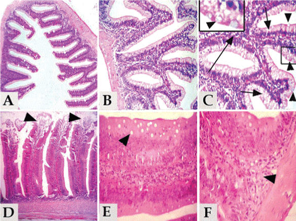
| Figure 3A: Section of Carassius auratus L. intestine of group 1, 2 and 3 showing normal, intact intestinal wall and intestinal villi. Figure 3B: Section of Carassius auratus L. intestine of group 1, 2 and 3 showing normal, intact intestinal mucosa, submucosa and muscularis. Figure 3C: Section of Carassius auratus L. intestine of group 1, 2 and 3 showing normal lining epithelium of simple columnar cells (short arrow) with scattered goblet cells (arrow head) and normal submucosa (long arrow).Inset box, showing higher magnification of the goblet cells. Figure 3D: Section of Carassius auratus L. intestine of group 4 (fasted group) showing necrosis in the intestinal villi with focal detachment of the lining epithelial (arrow head). Figure 3E: Section of Carassius auratus L. intestine of group 4 (fasted group) showing severe vacuolations of the cytoplasm of the intestinal columnar cells (arrow head). Figure 3F: Section of Carassius auratus L. intestine of group 4 (fasted group) showing chronic enteritis characterized by aggregation of mononuclear cells mainly lymphocytes, macrophages and plasma cell as well as fibrous connective tissue proliferation within the submucosa. |