|
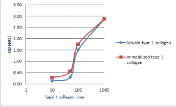
| Figure 1A: Binding of BP26 protein to type I collagen. Microtitreplates wells were coated overnight at +4°C with either BP26 (100 μg/ml in 2% BSA/PBS) or increasing concentrations of type I collagen (1000, 100, 50 and 10 μg/ ml in PBS-T. For negative controls, the wells were coated with 2% BSA/PBS instead of BP26 protein or collagen. Collagen coated wells (red bar) were incubated overnight at 4°C with 100 μl of Bp26 protein (100 μg/ml in 2% BSA/ PBS). Bound BP26 was detected with anti-BP26 mouse antiserum, followed by rabbit anti- mouse peroxidase conjugate and substrate. Bp26-coated wells (blue bar) were incubated overnight at 4°C with 100 μl of different concentrations of type I collagen (1000, 100, 50, and 10 μg/ml PBS/T), followed by incubation for 1 hr at 37°C with 1:1000 dilution of anti- type I collagen monoclonal antibody. Bound collagen was detected the reaction was visualized as above. Data was expressed as mean value of triplicates after subtracting negative control values. |