|
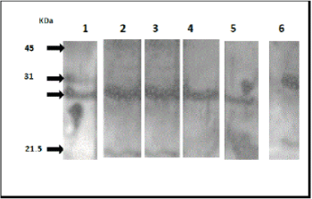
| Figure 1B: Collagen affinity blotting. BP26 was probed with anti- BP26 mouse antiserum (lane 1) and different concentrations of type I collagen (1000, 100, 50, and 10 μg/ ml then probed with anti-type I collagen monoclonal antibody (lane 3-6). Lane 2 represents a control where anti-type I collagen monoclonal antibody was omitted. Arrows indicate the position of BP26 bands. |