 |
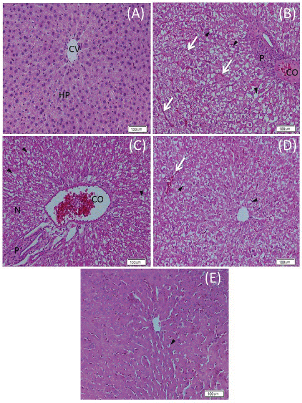
| Figure 4: Photomicrographs of liver sections of different treatment groups stained by H & E. (A) Control group showing normal histological structure of the hepatic parenchyma (HP) without any fibrosis or cirrhosis around central vein (CV). (B and C) Lornoxicam-treated group showing dilatation and congestion of blood sinusoids (arrows), central veins and portal blood vessels (CO), diffuse vacuolar degeneration (head arrows), focal hepatic necrosis (N) along with lymphocytic infiltration and proleiferation of bile duct (P). (D) LOR+ Basil oil-treated group showing mild hepatocytic vacuolar degeneration (head arrows) and few congested blood sinusoids (arrow). (E) LOR+ M. oleifera -treated group showing mild vacuolar degeneration (head arrows) with absence of fibrous tissue proleiferation. |