|
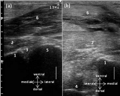
| Figure 2: Transcutaneous ultrasonograms of a transverse section of the ventral hip joint showing (a) the normal appearance and (b) the contralateral acetabulum without the corresponding femoral head in caudodorsal coxofemoral luxation. 1 Acetabulum, 2 Adductor muscle, 3 CJ coxofemoral joint, 4 Fossa acetabuli, 5 Femoral head, 6 Semimembranous muscle. Bar = 2 cm. |