|
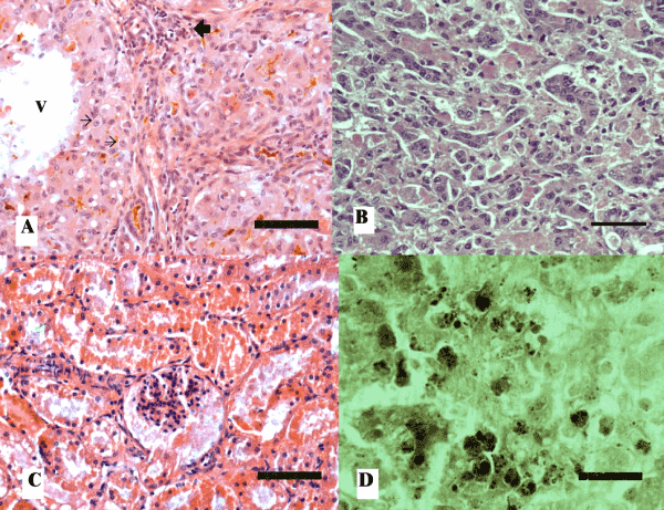
| Figure 1: A) Liver section from case N° 2 in which bile duct proliferation and fibrosis with mononuclear infiltration can be seen (large arrow). Towards the mid-zone and the hepatic vein (V), vacuolar changes and megalocytosis (small arrows) are evident. HE, bar= 50µm. B) Liver section from case N° 1, displaying sinusoidal disarrangement, and abundant cells exhibiting neoplastic changes such as altered nucleo-cytoplasm ratio and abnormal nuclear shape and size. Degenerative cells exhibiting nuclear pyknosis can also be seen. HE, bar= 50μm. C) Kidney section from calf N° 1 exhibiting tubulorrhexis with abundant strongly acidophilic material, resembling hemoglobin. Proteinaceous material is also evident in the capsule of the glomeruli. HE, bar= 50µm. D) Liver section from case Nº 1 in which strongly dark copper deposits can be seen in the cytoplasm of many cells. Timm’s stain, bar= 25µm. |