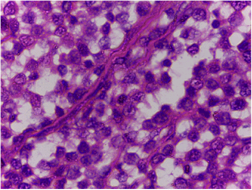
Figure 2:
Apocrine excretory adenoma; 2.5-year-old male, Great Dane dog X 100, H & E. Note multiple nodules of neoplastic cells which had a moderate amount of eosinophilic cytoplasm , separated by fibrovascular stroma.