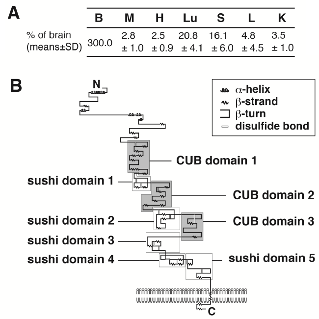
(A) Tissue distribution of Aig1l gene as detected by real-time quantitative PCR analysis (n = 5).B, Brain; M, muscle; H, heart; Lu, lung; S, spleen; L, liver and K, kidney.
(B) The secondary structure of Aig1l protein predicted by Pfam database and Jpred program. Aig1l protein is predicted to be a one-pass transmembrane protein, which has three CUB and five sushi domains.