|
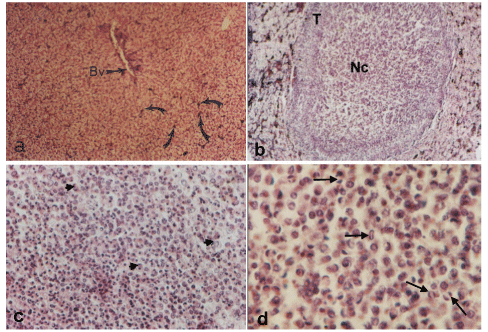
| Figure 1a-d: Light micrographs of sections of toads liver (Haematoxylin and Eosin stain): a- showing the architecture of normal liver. Hepatocytes are arranged in closely packed hepatic acini.Arrows point at pigment granules. Bv: blood vessel. (X 100). b- showing tumor nodule (T) in the liver of fluconazoletreated toad. Nc: Necrotic cells. (X 100). c- showing a high power view of focal liver lesion. Note large vesicular nuclei (arrows heads) and prominent nucleoli of tumor cells. (X 400). d- showing tumor cells with pleomorphic, irregular vesicular nuclei with prominent nucleoli. Arrows point at multiple round and abnormal mitotic figures. (X 1000). |