 |
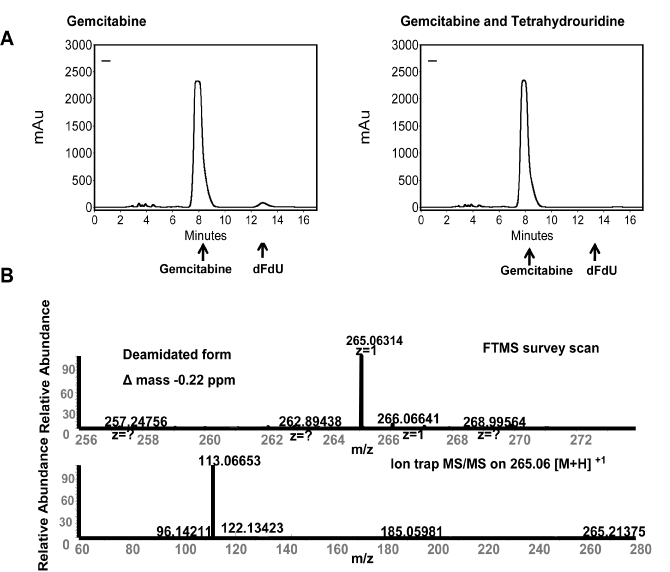
| Figure 1: HPLC and LC-MS/MS efflux analyses in SU-86 cells. (A) HPLC analyses of the extracellular media after gemcitabine treatment revealed two peaks with retention times of 8 min and 13 min, corresponding to gemcitabine and dFdU standards, respectively (left). The second peak disappeared after pretreatment with the deamination inhibitor, THU (right) confirming that is was dFdU. (B) Tandem mass spectrometry showed that the second retention peak had a precurser mass of 265.06314, and MS/MS spectra showed the same dominant 113.06 Da mass which corresponds to the 4-hydroxy-pyrimidin-2-one [M+H] +1 fragment, confirming that the second peak was dFdU. |