|
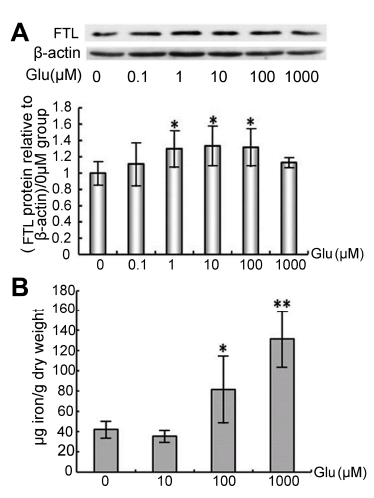
| Figure 3: The iron levels were increased in PC12 cells treated with different concentrations of Glu. A shows the expression of FTL in PC12 cells determined by western blot, and the histogram was the statistical relative values of FTL protein relative to control groups (0 μg Glu) after normalization by β-actin. B shows the total iron contents detected by ICP-MS in PC12 cells incubated with 0, 10, 100 and 1000 μM of Glu. All Data were expressed as means ± SD. * p<0.05 vs. 0 μg Glu, **p<0.01 vs. 0 μg Glu (n=6). |