|
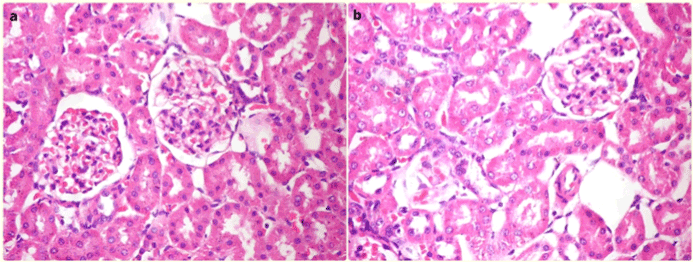
| Figure 2: Histological findings. a) Photomicrograph of irradiated group showing normal histological structure of glomeruli, renal tubules and interstitial tissue comprising renal cortex (H&E, X400). b) Photomicrograph of control group showing normal histological structure of renal cortex (H&E, X400). |