 |
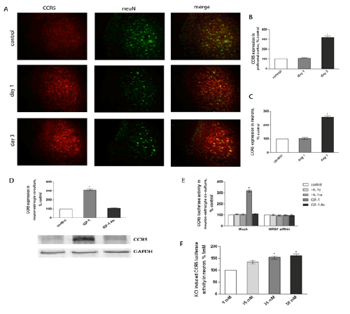
| Figure 4: CCR5 expression during traumatic stress. SD rats were divided into three groups: control, day 1, and day 3 (1 and 3 days after trauma; n=5). Cross sections of frontal cortex were subsequently immuno-stained by anti-CCR5 and Alexa594 antibodies, and anti-tubulin and Alexa488 antibodies, and were analyzed by Leika Q500IW image analysis system. Scale bars, 50 μm (A). Panels B and C show quantitative analysis of CCR5 expression, and co-localization of CCR5 with neuN, respectively. Neurons and astrocytes were separated from prefrontal cortex and cultured for the indicated times: (D) in neurons, CCR5 activity in the presence of 5–50 mM of KCl was detected by luciferase assay; (E) in co-culture of neurons and astrocytes, IGF-1 induced CCR5 expression was measured by western blot analysis; (F) in co-culture of neurons and astrocytes, after transfection of NRSF or NRSF siRNA, CCR5 activity was measured by luciferase assay. Data were calculated as percentage of control; each value represents the mean ± SEM of three independent experiments. *p<0.05 vs. control or 5 mM. |