 |
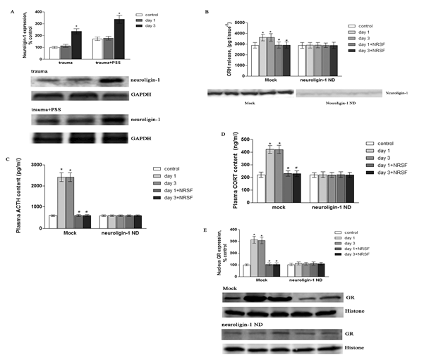
| Figure 9: Contribution of NRSF in the altered HPA axis is dependent on neuron-astrocyte communication. SD rats underwent traumatic stress, were subjected to predator scent exposure following trauma (trauma+PSS) for 14 days, the prefrontal cortex was collected and homogenized, and neuroligin-1 expression was determined by western blot analysis (A). SD rats were infected with Mock or negative dominant neuroligin-1, then exposed to traumatic stress, or subjected to predator scent exposure following trauma (trauma+PSS) for 14 days, and NRSF was over-expressed by adeno-virus: (B) hypothalamus was sonicated using a tissue extraction reagent supplemented with a protease inhibitor cocktail and the homogenate was centrifuged (10 min, 14,000 g, 4°C) and the supernatant collected; (C and D) cardiac blood was centrifuged (10 min, 14,000 g, 4°C), and CRH release, ACTH and CORT were measured using a competitive immunoassay (Assay Designs, Inc., Ann Arbor, MI), in this part, animals exposed to PSS but not undergone traumatic stress were used as control group. Total protein was quantified using a Bradford assay. (E) GR expression in the nucleus was measured by western blot analysis. Data were calculated as percentage of control; each value represents the mean ± SEM of three independent experiments. *p<0.05 vs. control, #p<0.05 vs. day 1/3. |