 |
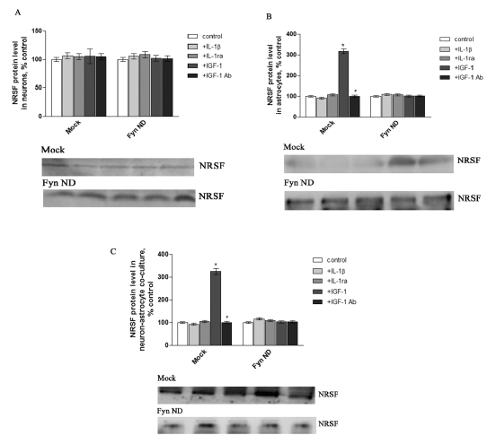
| Figure 2: Neuron-astrocyte communication dependent NRSF expression. Neurons and astrocytes were separated from prefrontal cortex and cultured for the indicated times, then transfected with Mock or negative dominant Fyn; 48 h later, cells were exposed to IL-1β/IL-1ra or IGF-1/IGF-1Ab. NRSF expression in neurons (A), astrocytes (B), and co-culture of neurons and astrocytes (C) was determined by western blot analysis. Data were calculated as percentage of control; each value represents the mean ± SEM of three independent experiments. *p<0.05 vs. control, #p<0.05 vs. IGF-1. |