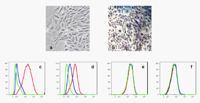
Growing MSC develop as a monolayer of semi- confluent cells (a) which becomes confluent after 14-16 days in culture (b).
After releasing confluent MSC from the tissue culture flasks, a partial immunophenotype was assessed by Flow Cytometry. Histograms show: c) CD44 (90%); d) β1 Integrin (85%); e) CD34 (0%); f) CD45 (0%).
Values in parenthesis indicate the percentage of expression of each antigen. Lines in the histogram correspond to autofluorescence (green), secondary antibody (blue) and primary antibody (red).