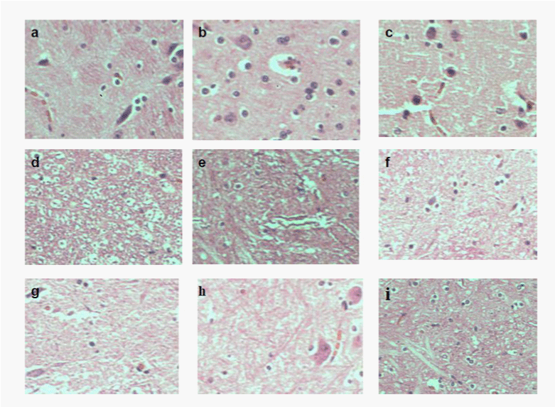
Hemotoxylin and eosin-stained sections prepared from a rabbit infused with a high dose of MSC. Pictures correspond to: hindbrain (a); brainstem (b); cerebellum (c); upper cervical cord (d); mid cervical cord (e) and serial sections of lumbar sacral cord (f-i).