|
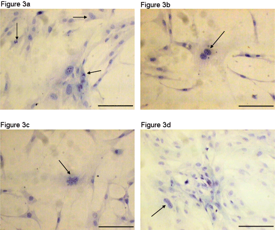
| Figure 3: Pap test results, Sample I, human AT-MSCs. Papanicolaou stain, P2, 100× optical microscopy. (a) Cells with atypical mitosis (arrows), (b) cell with an increased number of nuclei (arrow), (c) Cell with an increased number of nuclei (arrow) and (d) cell with an atypical and multilobulated nucleus (arrow). The analysis result according to the Bethesda classification was high-grade alterations consistent with genetic instability. Scale bars = 10μm. |