|
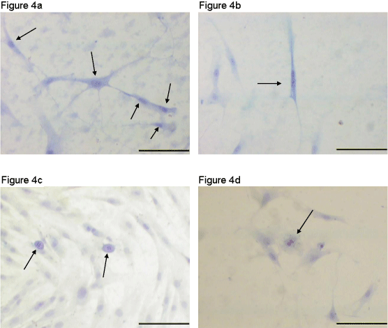
| Figure 4: Pap test results, human AT-MSCs. Papanicolaou stain, P2, 100 X optical microscopy. (a) Sample III, cells with normal morphological aspects and (b) cell with a double nucleus (arrow). The analysis result according to the Bethesda classification was atypical cells of undetermined significance. (c) Sample VI, cells with normal morphological aspects. Typical mitosis in telophase (arrow) and (d) Cells with normal morphological aspects. Typical mitosis in anaphase (arrow). The analysis result according to the Bethesda classification was normal cells. Scale bars = 10μm. |