 |
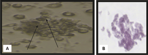
| Figure 1: Morphological and histological staining of differentiated BM-MSCs into osteoblasts. (A) (×20) Arrows for differentiated MSCs osteoblasts after addition of growth factors. (B) (×200) Differentiated MSCs into osteoblasts stained with Alizarin red stain.. |