|
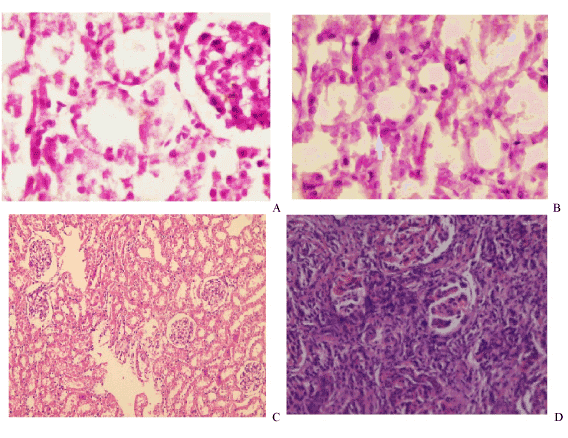
| Figure 7: Histopathological examination of renal tissues in different groups: (A)ARF showed atrophy and patchy necrosis of proximal and distal renal tubules & cell debris in the lumen. (B)ARF+ MSC(B) & ARF+MSC+HGF(D) showing dense interstitial tissue infiltrate between tubules at corticomedullary junction.(D)ARF+HGF (C), showed fibrin and cell debris in cortical tubules. |