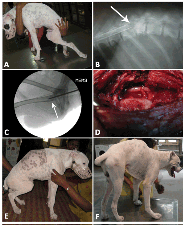
Legend: (A) Pre-operative Grade IV Paraplegia, (B) Pre-operative Myelogram in which the 12th vertebral compression can be visualized (See arrow), (C) C-Arm image of Bone Marrow Harvesting;the arrow indicates tip of the Jamshidi needle inside the Bone Marrow, (D) intra-operative image showing exposed spinal cord after hemilaminectomy in which the spinal cord is observed to be odematous and bulged out with intact duramater, (E) Post-operative Day 53 on which transient unassisted standing on the hind limbs by the canine was observed (F) Post-operative Day 98: The Canine could move around with incoordinated left hind limb movements.