|
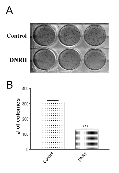
| Figure 2: Autocrine TGF-ß signaling supports anchorage independent growth. (A). Exponentially growing cells were suspended in 1 ml 0.4% soft agarose in 10% FBS containing medium and plated on top of a 1 ml solidified underlayer of 0.8% agarose in the same medium in a 6-well tissue culture plate. After 2 weeks of incubation, cell colonies were visualized by staining with 1 ml p-iodonitrotetrazolium violet. (B) The stained dishes were digitally scanned and the colonies that were greater than 5 pixels were manually counted after the scanned pictures were displayed in PhotoShop (***P<0.001). |