|
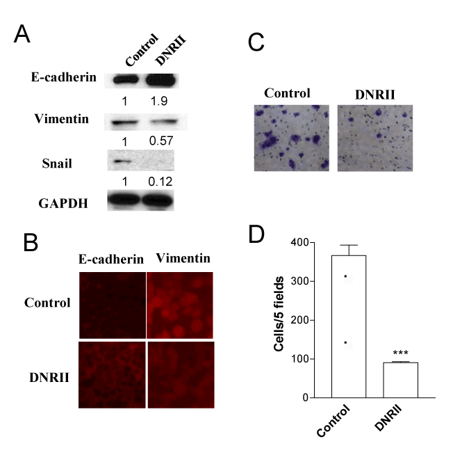
| Figure 4: Autocrine TGF-ß signaling is necessary for the progression of EMT in NMuMG-ST cells. (A) Cells from exponential cultures of the control and DNRII cells were plated in T-25 flasks. After culturing for 4 days, cell extracts were used for Western blot analysis to detect the expression of E-cadherin, Vimentin and Snail. The density of each band was quantified with The Image J software and divided by the density of the corresponding GAPDH band. The ratio is presented under each blot after normalizing the values for control as one unit. (B) Control and DNRII cells were grown on cover slips in a 24-well plate till 80% confluence. Cells were fixed, permeabilized, blocked and incubated with an anti- E-cadherin or anti-vimentin antibody followed by the incubation with fluorescent dye-tagged secondary antibody. (C) and (D). In a Boyden chamber, 25,000 cells in serum-free medium were seeded in the upper chamber. The lower chamber contained the medium with 10% of FBS as chemoattractant. After incubation for 16 hours, cells were removed from the upper surface of the chamber membrane and stained for the migratory cells at the lower surface of the chamber with HEMA3 staining solution. Cells were counted under a microscope (***p<0.005). |