|
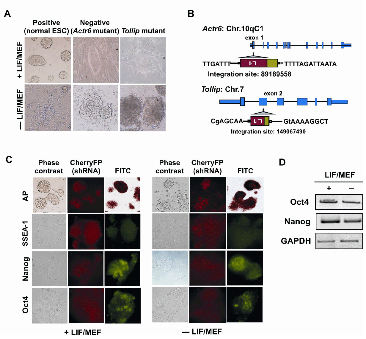
| Figure 3: Verifying the loss of the cell differentiation ability. (A) Inverted microscopic image of ES cells harboring an L1 insertion. Normal ES cells and Actr6-disrupted ES cells (A2 clone) were used as the positive and negative controls, respectively, for cell differentiation. Tollip-disrupted ES cells (B1 clone) completely failed to differentiate in the absence of LIF even after 4 weeks. Plus, presence; minus, absence of LIF and MEFs. (B) The identification of disrupted Actr6 and Tollip genes by inverse PCR. The disrupted sites from the L1 insertions and their positions with regard to the exon-intron organization of the genes are shown. (C) Validation of the Tollip mutation after treatment with Tollipspecific shRNAs in the normal ES cells. Immunofluorescence staining of the undifferentiated versus the differentiated Tollip-knockdown ES cells are shown. Note the loss of differentiation without LIF/MEFs. (D) Quantification of endogenous Oct4, Nanog and GAPDH (internal control). qRT-PCR analysis performed in the presence or absence of LIF/ MEFs after Tollip-specific shRNA transfection in the wild-type ES cells. Plus, presence; minus, absence of LIF and MEFs. |