|
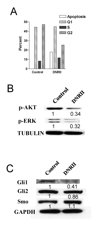
| Figure 3: Abrogation of autocrine TGF-ß signaling induces apoptosis and downregulates cell survival pathways. (A) Control and DNRII cells were plated at 0.4 x 10⁶ in a 60mm dish. After the culture reached confluency, the culture medium was switched to a serum free medium for 4 days. The cells were fixed with ethanol, stained with propidium iodide and analyzed by Flow Cytometry. Percent of cells with DNA content consistent with various cell cycle or apoptosis stages are plotted. (B) and (C) Western blots for the p-AKT, p-ERK, Gli1, Gli2 and Smo expression in control and DNRII cells. The density of each band was quantified with the Image J software and divided by the corresponding density of Tubulin or GAPDH band. The ratio is presented under each blot after normalizing the values for control as one unit. |