 |
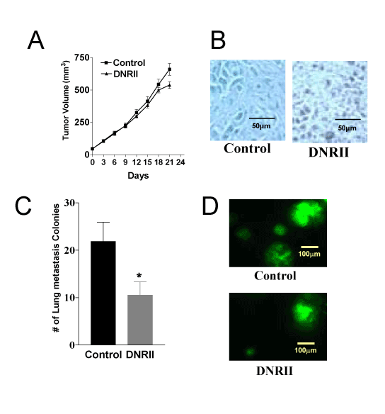
| Figure 6: Blockade of autocrine TGF-ß signaling induces apoptosis and inhibit lung metastasis in vivo. (A) Cells from exponential cultures of the control and DNRII cells were inoculated subcutaneously in the flanks of 5-week-old female athymic nude mice. The growth of tumors was monitored every three days and tumor sizes were measured with a caliper in two dimensions. Tumor volumes were calculated with the equation V= (LxW2) x 0.5, where L is length and W is width of tumor. The data are presented as mean+SEM of 14 tumors in the control group and 12 tumors in the DNRII group. (B) Tumors formed by the control and DNRII cells in nude mice were fixed in buffered formalin, embedded in paraffin and 5 mm sections were used for TUNEL staining. The cells stained in brown color are apoptotic. (C) Lungs were removed during autopsy and metastasis colonies expressing GFP in each mouse lung were counted under TE-200 Nikon fluorescence microscope. The data are presented as mean+SEM of lung nodules from 6 mice in each group (*p<0.05). (D) Representative pictures of lung metastasis colonies (x40 magnification) formed by the control and DNRII cells. |